From an mRNA Display Hit to Enlicitide (2): Seeing How the Macrocycle Binds PCSK9 for the First Time
Compound 30 enabled the first co-crystal structure for this PCSK9 macrocycle series, making its binding mode visible and turning empirical SAR into testable structure-based design hypotheses.
From an mRNA Display Hit to Enlicitide (2): Seeing How the Macrocycle Binds PCSK9 for the First Time
From an mRNA Display Hit to Enlicitide · Part 2
Previous: The Story Begins with a Screen
Why Can One Crystal Structure Change a Drug-Discovery Program?
The first article in this series stopped at a familiar but uncomfortable point in peptide discovery. An mRNA display screen had found cyclic peptides capable of binding PCSK9, and early medicinal chemistry had made the selected scaffold smaller and more potent. Yet the team was still working with indirect evidence. A substitution could improve activity, another could damage it, and truncation could reveal that part of the original hit was unnecessary—but an activity result alone could not show why.
Compound 30 marked a different kind of step forward. It was not important only because it represented another potency improvement. It enabled the first co-crystal structure for this cyclic-peptide series, turning a pattern of empirical structure–activity relationships into a visible molecular binding mode.
The structure deposited as PDB 6XIB contains an 11-residue macrocyclic peptide bound to PCSK9. Its sequence includes two 5-fluorotryptophan residues and an α-methylproline, while two cysteine side chains are joined through the 1,3-di(bromomethyl)benzene-derived DBX bridge. Those details matter because the molecule in the crystal is not a generic peptide ring: its noncanonical chemistry and cyclization are part of the object being recognized by PCSK9.
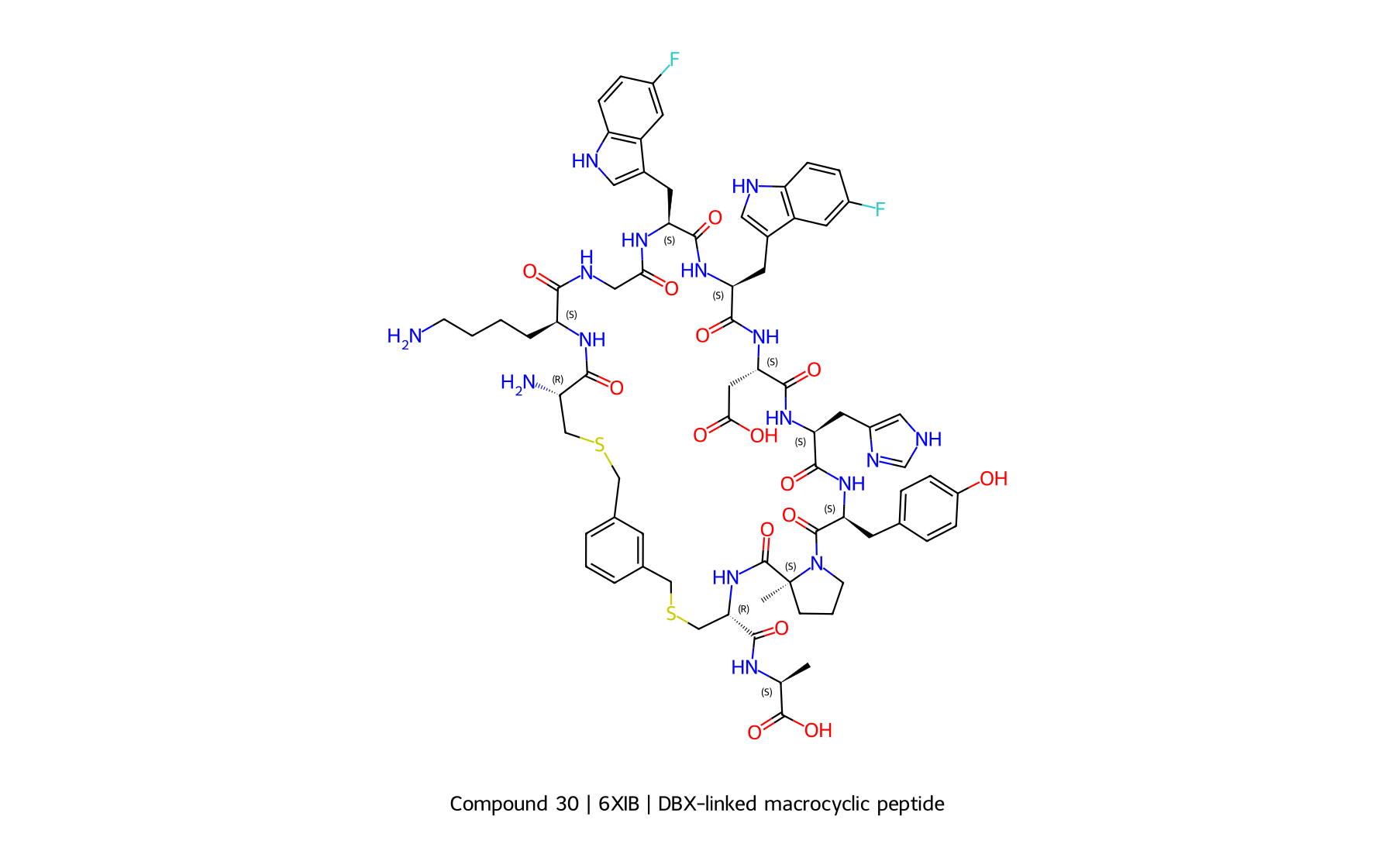
Figure 1. Compound 30: A Key Molecule in the Evolution of the PCSK9 Macrocycle Program. The structure was rebuilt with RDKit from the 6XIB polymer sequence, modified-residue definitions, and covalent-link records; it is not a screenshot of the publication figure. Sequence: Cys–Lys–Gly–5F-Trp–5F-Trp–Asp–His–Tyr–α-Me-Pro–Cys–Ala, with a DBX bridge between the two cysteine sulfurs.
From “Which Changes Work?” to “Why Do They Work?”
Before a reliable complex structure is available, medicinal chemistry often proceeds by controlled comparison. Change one residue, measure activity, and ask whether the molecule improved. Accumulated results can reveal useful SAR, but the interpretation remains incomplete. A loss of activity could reflect the removal of a direct target contact, disruption of the macrocycle’s preferred conformation, a change in solubility, or some combination of these effects.
A co-crystal structure does not eliminate experiments, but it changes the questions that experiments can answer. Residues can now be viewed in the context of the protein surface. A side chain that appeared important in an assay can be evaluated as a potential contact; a region that tolerates substitution may face solvent; and a residue with no obvious direct protein interaction may still help organize the macrocycle into its binding-competent shape.
This is the transition from empirical SAR to structure-based design. It does not mean that the structure predicts every successful change. It means that chemical modifications can be proposed with an explicit molecular hypothesis and tested against the next experiment. The same principle underlies careful structure-aware optimization of an existing peptide: computation and structural models help prioritize hypotheses, but measured binding and developability data remain the final test.
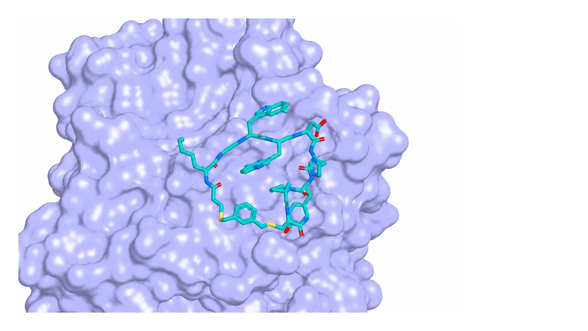
Figure 2. How Compound 30 Binds PCSK9. Original rendering from the 1.55 Å X-ray structure PDB 6XIB. PCSK9 is shown as a pale-blue surface and Compound 30 as sticks; crystallographic waters and glycerol are omitted for clarity. The figure uses deposited atomic coordinates and does not infer additional contacts.
Why Was a Noncanonical Amino Acid So Important?
The original mRNA display campaign already included 5-fluorotryptophan in the accessible amino-acid set. Compound 30 makes that design choice tangible: the deposited structure contains two 5F-Trp residues, and the binding mode places these large aromatic side chains in defined local environments on the PCSK9 surface.
Fluorination can look like a minor edit when written as a sequence annotation. In three dimensions, however, even a small chemical substitution can alter how a side chain occupies a local pocket, distributes electron density, or balances interactions with its surroundings. The important lesson is not that fluorination is universally beneficial. It is that a noncanonical amino acid can participate in highly specific molecular recognition, and its value depends on position, conformation, and target context.
This is also why adding noncanonical amino acids during library construction can be more powerful than treating them only as post-screen substitutions. Selection can evaluate the modified chemistry as part of the binding molecule itself. The first article’s discussion of mRNA display libraries containing noncanonical amino acids and cyclic peptides provides the broader discovery context.
The Macrocycle’s Shape Became as Important as Its Sequence
A peptide sequence is a one-dimensional description. The target encounters a three-dimensional object. For a macrocycle, that object is determined by the sequence, noncanonical chemistry, cyclization geometry, and the conformational preferences created by all of them together.
Compound 30 illustrates why this distinction matters. The DBX bridge does more than connect two cysteines on paper. It constrains the chain and helps define which arrangements are readily accessible. α-Me-Pro is likewise not simply a residue name inserted into the sequence. Its backbone substitution changes the local conformational landscape. These effects can position target-facing side chains without every constraining element needing to make a direct contact with PCSK9.
The crystal structure therefore made a second layer of SAR visible. Some changes affected recognition because they altered a contact; others could affect recognition by changing the shape that presents those contacts. Sequence, chemistry, conformation, and target interaction could no longer be interpreted independently.
The Structure Showed Where Change Might Be Possible
A useful structure identifies more than an immutable binding motif. It begins to separate regions that play different roles. Some parts of the macrocycle appear central to recognition and deserve conservative treatment. Others help maintain the bound conformation. Still others are more solvent exposed and may offer room to adjust molecular properties.
That distinction matters because the next problems were not limited to affinity. The program still needed to improve protease stability, metabolic behavior, solubility, permeability, pharmacokinetics, and ultimately oral exposure. A solvent-facing position might accept a change intended to tune solubility. A conformational position might be modified to reduce flexibility or shield a vulnerable backbone region. A target-facing residue, by contrast, could carry a much higher risk of losing binding.
None of those assignments should be treated as a guarantee. Crystal structures are snapshots, solution ensembles remain dynamic, and a chemically attractive design can fail for reasons not visible in one model. But the structure turns undirected changes into testable choices: preserve this interaction, restrain that conformation, and explore this exposed region for property optimization.
From One Structure, the Question Changed
mRNA display had answered, “What kind of cyclic peptide can bind PCSK9?” The first co-crystal structure began to answer, “Why can it bind?” From that point forward, the project could ask a more demanding question: how can the molecule be changed to become more stable and more drug-like without losing the recognition mode that made it valuable?
That was the real importance of Compound 30. It did not finish the optimization campaign, and it was not enlicitide. It gave medicinal chemists a molecular map for the next stage of the journey.
Previous: The Story Begins with a Screen
Next: Binding Well Is Not Enough
Structure-guided D-amino-acid substitution, additional cyclization, and N-methylation improved stability while exposing new PK, safety, and solubility liabilities.
Scientific Sources and Related Reading
Alleyne C, Amin RP, Bhatt B, et al. Series of Novel and Highly Potent Cyclic Peptide PCSK9 Inhibitors Derived from an mRNA Display Screen and Optimized via Structure-Based Design. Journal of Medicinal Chemistry. 2020. https://doi.org/10.1021/acs.jmedchem.0c01084
RCSB Protein Data Bank. PCSK9(deltaCRD) in complex with cyclic peptide 30. PDB 6XIB, X-ray diffraction, 1.55 Å. https://www.rcsb.org/structure/6XIB
Related services and resources: mRNA display technical resources | AI peptide design | custom peptide synthesis
Have a cyclic-peptide hit or structural data ready for the next optimization cycle?
Apollomics supports custom synthesis, structural confirmation, and multi-parameter experimental validation of noncanonical macrocycles, helping teams turn structural hypotheses into testable molecules.