When a Peptide “Disappears” in Plasma: How Can You Tell Whether It Was Proteolyzed?
A falling parent-peptide LC-MS signal can reflect adsorption, extraction recovery, matrix suppression, or true proteolysis. This article builds the evidence chain from controls and degradation products to the next sequence-design decision.
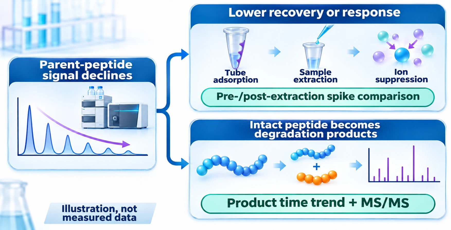
A peptide is incubated in plasma, and its parent LC–MS peak falls with time. It is tempting to call the result “enzymatic degradation,” but that conclusion is premature. The peptide may have been cleaved; it may also have adsorbed to a tube, been lost during extraction, or suffered stronger ion suppression in the matrix. The first task is therefore not to name an enzyme, but to separate true loss of intact peptide from loss during sample preparation or detection.
First establish that the parent peptide really decreased
Time zero and later samples should experience the same incubation stop, extraction, transfer, and LC–MS analysis. A buffer-only control can reveal whether the peptide is intrinsically unstable in that buffer, but it cannot reproduce plasma protein binding, extraction loss, or matrix ion suppression and therefore cannot by itself prove enzymatic degradation.
A practical way to separate these effects is to compare spikes made before extraction with otherwise matched spikes made after extraction. Their difference estimates extraction recovery. Comparing a post-extraction spike in extracted blank matrix with the same amount in neat solution estimates the matrix effect on ionization. This is the logic behind the three-set framework described by Matuszewski and colleagues; the exact concentrations and replicates still need to be designed for the candidate and method.
An intact stable-isotope-labeled version of the peptide is especially useful because it can track many losses and response changes that affect the analyte similarly. Timing matters, however. An internal standard added only after extraction cannot correct adsorption or poor recovery that occurred earlier, and one added before plasma incubation may itself undergo degradation. The standard should therefore be added at the stage appropriate to the question, with its own stability verified.
Turn signal loss into evidence of cleavage
A stronger degradation claim combines two time trends: the intact parent falls while one or more chemically plausible products rise. LC–HRMS can generate candidate product formulas from accurate mass, but accurate mass alone is not a cleavage-site assignment. Oxidation, deamidation, adducts, isotope selection errors, and in-source fragmentation can all create misleading features. Product-ion spectra should support the proposed sequence fragments; when the decision is important, retention time and MS/MS can be compared with a synthesized product standard.
For a simple head-to-tail cyclic peptide, the first hydrolytic ring opening adds one water molecule, giving a theoretical neutral-mass increase of 18.0106 Da. That mass shift is a useful search clue, not proof: retention behavior and fragmentation are still needed to distinguish the opened product from alternatives. Conversely, failure to observe a product does not prove that cleavage did not occur. The first product may be degraded again, ionize poorly, adsorb, or lie outside the acquisition and data-processing window.
Peak areas from different peptide species should not be added as though they formed a mass balance. Sequence changes can alter recovery, charge-state distribution, chromatography, and ionization by orders of magnitude. Quantitative comparison requires a response model or an appropriate standard for each product. Enzyme attribution should be graded in the same way: a plausible motif is a hypothesis; selective inhibition, purified-enzyme reproduction, product mapping, and loss of the effect under a targeted perturbation provide progressively stronger evidence.
What Compound 32 in the zilucoplan story does—and does not—show
In the zilucoplan discovery paper, Compound 32 retained about 95% after 24 hours in human plasma but about 2% in rat plasma in an in-vitro, single-time-point experiment. The authors attributed the difference to species-specific enzymatic degradation. This is valuable evidence of a species-dependent stability liability, but it should not be extended beyond the reported experiment: it does not identify the responsible enzyme, provide a complete degradation curve, or establish in-vivo pharmacokinetics.
Plasma stability and pharmacokinetics answer different questions. An intact peptide can be stable during an in-vitro plasma incubation and still be cleared rapidly in vivo through renal filtration, tissue uptake, or other routes. Conversely, a plasma-stability problem can be masked in exposure data by distribution or formulation effects. The sequence-optimization context for Compound 32 is described in From an mRNA Display Hit to Zilucoplan (2); broader distinctions among proteolysis, clearance, release, and formulation are discussed in Four Questions Behind Peptide Stability.
Translate a cleavage map into the next synthesis
The next analogue should respond to the observed site rather than receive every available stability modification. Predominantly terminal truncation makes terminal capping or a topology that removes the accessible terminus a logical test. A defined internal cleavage site supports local substitution with a D- or noncanonical amino acid, backbone modification, or a conformational constraint around that site. Each intervention should retain the original parent as a control.
The comparison then returns to two linked readouts: how much intact parent remains, and whether target activity is retained. Product mapping should also be repeated, because blocking one site may reveal a second cleavage pathway. This cycle—measure the parent, verify the product, modify locally, and retest activity—turns a disappearing LC–MS peak into a defensible sequence-design decision.
References
- Ye P, Hammer RP, Wang Z, et al. Discovery of Zilucoplan: A Potent Macrocyclic Peptide Inhibitor of Complement Component 5. Journal of Medicinal Chemistry. 2025;68:25772–25782. https://doi.org/10.1021/acs.jmedchem.5c02537
- Matuszewski BK, Constanzer ML, Chavez-Eng CM. Strategies for the Assessment of Matrix Effect in Quantitative Bioanalytical Methods Based on HPLC–MS/MS. Analytical Chemistry. 2003;75(13):3019–3030. https://doi.org/10.1021/ac020361s
- Hoofnagle AN, Whiteaker JR, Carr SA, et al. Recommendations for the Generation, Quantification, Storage, and Handling of Peptides Used for Mass Spectrometry-Based Assays. Clinical Chemistry. 2016;62(1):48–69. https://doi.org/10.1373/clinchem.2015.250563