From an mRNA Display Hit to Zilucoplan (2): How Were Two Hits Combined into a More Stable Macrocycle?
Compound 1 was more potent but its linear structure exposed plasma-stability problems; Compound 2 was somewhat less potent but already macrocyclic. The team combined features from both to obtain Compound 32, then continued by optimizing the cyclization mode and degradation-prone sites.
From an mRNA Display Hit to Zilucoplan · Part 2
← Previous: Two screening hits, two different clues
Why Was the More Potent One Still Not Enough?
In the previous article, the research team obtained two C5 inhibitor starting points through mRNA display screening. Compound 1 was more potent, with an IC₅₀ of 12 nM in a red-blood-cell lysis assay. Compound 2 had a somewhat weaker IC₅₀ of 27 nM, but it had already been cyclized through two cysteines and a xylene-derived linker. These IC₅₀ values came from a functional C5 inhibition assay; they are not measurements of molecular binding affinity to C5.
The linear structure of Compound 1 created two specific problems: the cysteine at position 2 was susceptible to oxidation and dimer formation, while phenylglycine at position 12 was prone to epimerization under standard coupling and deprotection conditions, producing an isomer that was difficult to separate and characterize. Plasma-stability experiments also showed that, after 24 hours of incubation, only about 9% of Compound 1 remained in human plasma and about 4% remained in mouse plasma. Attempts to cyclize it directly were unsuccessful. The researchers therefore faced more than the question of how to improve potency: they also needed to change the structure so that the molecule could persist more stably.
Combining Features from the Two Hits
Compound 2 supplied a cyclic structure that could be used. The research team combined its N-terminal cyclic portion with the C-terminal portion of Compound 1 to design the 15-amino-acid Compound 32. It retained strong activity, with an IC₅₀ of 9 nM in the red-blood-cell lysis assay; after 24 hours of incubation in human plasma, approximately 95% of the molecule remained intact. Compared with Compound 1, this result showed a marked improvement in stability.
This modification did not eliminate every problem at once. After 24 hours of incubation in rat plasma, only about 2% of Compound 32 remained. The paper's authors suggested that this might be related to species-dependent differences in enzymatic degradation. The result also reminds us that in vitro plasma stability cannot be equated directly with in vivo pharmacokinetic performance: a molecule that is stable in human samples will not necessarily behave the same way in other species or in an in vivo environment.

Figure 1. Compound 32 combines the cyclic head of Compound 2 with the tail of Compound 1. Colors indicate structural origin and positions requiring attention, not validated C5 binding sites.
After Stability Improved, New Degradation Sites Appeared
The research team next continued to adjust the mode of cyclization. After a series of comparisons, they selected Compound 37, which forms a lactam ring between lysine and aspartic acid. This design retained strong activity in the red-blood-cell lysis assay, with an IC₅₀ of approximately 5 nM; using a lactam linkage also helped avoid possible redox problems associated with sulfur-containing linkages.
Metabolite analysis found that a protease cleavage site remained near the exocyclic aspartic acid in Compound 37, and its C-terminal amide also underwent deamidation. The researchers therefore introduced N-methyl aspartic acid at position 7 and changed the C-terminal amide to a carboxylic acid, producing Compound 38. Its IC₅₀ decreased to 0.9 nM; after 4 hours of incubation, approximately 84%, 62%, and 88% remained in human, monkey, and rat plasma, respectively. This step both increased functional inhibitory potency and made specific changes aimed at the degradation problems identified in the preceding step.
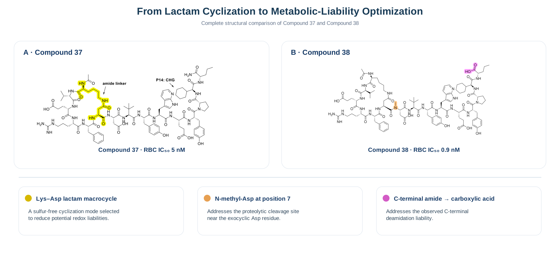
Figure 2. Structural comparison of Compound 37 and Compound 38. The lactam ring, N-methyl aspartic acid at position 7, and the change from a C-terminal amide to a carboxylic acid are highlighted. These structural changes correspond, respectively, to selection of the cyclization mode and optimization of metabolic liabilities.
Compound 38 was an important stage for in-depth characterization, but it cannot be equated directly with the final drug zilucoplan. A subsequent intravenous study in monkeys measured a half-life of approximately 20.7 hours, providing a basis for continuing toward the goal of once-daily subcutaneous administration. The next article will continue from here: how the research team further adjusted in vivo exposure through lipidation, albumin binding, and spacer design.
Next in the series: How Was a Macrocyclic Peptide Turned into a Once-Daily Subcutaneous Drug?
Scientific Source
Ye P, Hammer RP, Wang Z, et al. Discovery of Zilucoplan: A Complement C5 Inhibitor for Treatment of Anti-Acetylcholine Receptor (AChR) Antibody-Positive Generalized Myasthenia Gravis (gMG). Journal of Medicinal Chemistry. 2025;68(24):25772–25782. https://doi.org/10.1021/acs.jmedchem.5c02537